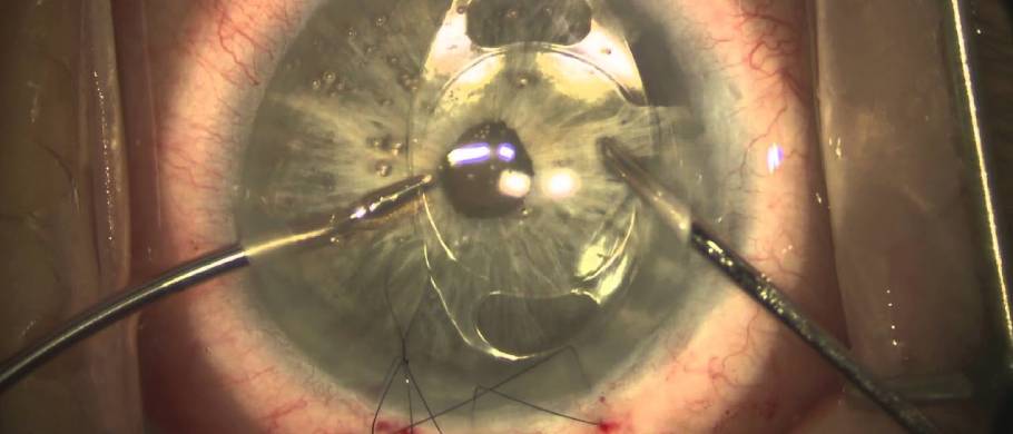
Secondary IOL Surgery
Secondary intraocular lens (IOL) implantation has evolved over the past few decades. Several new techniques, lens options, and materials now exist. Careful patient selection is important to determine the optimal secondary IOL technique. Intraocular lens placement in the capsular bag is the most ideal followed by sulcus placement. However, the best option when no capsular support exists in an aphakic patient remains unclear. Surgeons should be aware of contraindications for each technique; however, there are several situations where anterior chamber intraocular lens (ACIOL), scleral-fixated intraocular lens (SFIOL), and iris fixation can all be used. In those cases, surgeon familiarity and comfort with the secondary IOL technique can determine the type of surgery performed
Introduction
Secondary intraocular lens implantation is defined as implantation of an intraocular lens following an initial surgery that resulted in aphakia or a deficient intraocular lens. The indications for secondary intraocular lens insertion have evolved with improved surgical outcomes of modern cataract surgery. Newer surgical techniques and lenses have also advanced the field of secondary intraocular lenses. The first wave of secondary intraocular lenses to be implanted was the anterior chamber intraocular lens (ACIOL). Secondary intraocular lenses can now be implanted in a variety of anatomic locations with different techniques used to support the lens (sutured, iris-claw, etc.). Specifically, sutured IOL and intrascleral fixation techniques have been gaining popularity. From the statistics it has been found that 538% increase in secondary sutured IOL techniques from 2000 to 2013. However, with the advent of several new techniques, there is no clear guidance for the best technique for secondary IOL placement. This chapter aims to discuss the variety of secondary intraocular lenses, the indications for use, and surgical considerations
Indication
Modern cataract surgery has evolved the role of secondary intraocular lens implantation since there is now less incidence of surgical aphakia after cataract surgery. With current technology and improved cataract surgery technique, the most common reason for secondary lens implantation is IOL exchange. The rates of IOL exchange also have declined over the years with recent studies showing rates of 0.34–0.77%. ACIOL explantation is most commonly due to corneal decompensation and inflammation. PCIOL explantation is most commonly due to IOL decentration and dislocation. IOL dislocation can be due to zonular dehiscence from trauma, previous complicated surgery, or conditions predisposing to zonular instability such as pseudoexfoliation syndrome and Marfan’s syndrome.
In recent years, advancements in IOL calculations, cataract surgery technology and technique have improved refractive outcomes. Patient visual expectations after cataract surgery have increased and now, in some cases, IOL exchanges are performed for unexpected refractive outcomes, dissatisfaction with multifocal lenses, and dysphotopsias following cataract surgery. The rates of IOL exchange due to patient dissatisfaction in one study showed an increase from 7.8% in 2005 to 21% in 2014. In 2005, no patients underwent IOL exchange for unsatisfactory refractive outcomes in the absence of optical aberrations but in 2014, 42% of IOL exchanges were due to unsatisfactory refractive outcomes alone.
Preoperative Evaluation
Prior to consideration of secondary intraocular lens implantation, a thorough pre-operative history is required. In particular, details of the prior cataract removal including intraoperative complications, type of IOL implanted, location of the IOL implant and the presence of other ocular hardware including glaucoma drainage devices are important pieces of information to gather before secondary IOL surgery. To this end, review of prior operative reports and medical records is a critical element of every preoperative evaluation.
A thorough examination of the anterior and posterior segment is required to plan for a secondary IOL implantation. The conjunctiva and scleral should be examined to identify any prior incisional glaucoma surgery or devices. Corneal health should be evaluated to determine if an ACIOL is a viable option. Specular microscopy or pachymetry can be obtained as needed to assess corneal endothelial health. Anterior chamber depth should be evaluated as a narrow/shallow chamber might preclude safe ACIOL placement.
The presence of vitreous prolapse in the anterior chamber should be noted as well as the integrity of the iris and capsule. Of note, high-frequency ultrasound has shown to be better than slit-lamp examination in assessing capsular support for sulcus IOL implantation. If there is an intraocular lens in place, the type of lens and degree of dislocation should be assessed. The optic nerve and retina should be thoroughly examined to evaluate for any other ocular comorbidities that can limit vision potential or require treatment at the time of secondary IOL implantation. Finally, vision potential with reliable manifest refraction is important to gauge the potential benefit of secondary IOL implantation
How To Reach Us?
Dr. Nilesh Giri is a trusted Eye Specialist in PCMC, Pune. At Devgiri Memorial Hospital you will get all the necessary medical treatment. Our advanced approach for our patient treatments make us unique.
The appointment process at Devgiri Memorial Hospital PCMC Pune is very simple. You can directly call on 09657002695. Also with the help of the “Book an Appointment” Form you can book your appointment by just filling in the basic information. We will contact you via email or phone call to confirm your appointment.